Advanced Dental Technology
Welcome to the 21st Century dentistry at World Dental Specialities in Mumbai.
Today, more than ever, dentistry is a dynamic field, changing very, very quickly. We are always on the top of any technology that can help us achieve our target successfully time after time. Our patients appreciate our efforts toward this paradigm shift in dental technology only to improve and cross all barriers towards excellence in dentistry. This finally reflects in our work and their results!
This is our commitment of excellence at World Dental Specialities, where your comfort and pioneering dentistry come hand in hand. We also pledge to involve you in the decision-making process so that you are aware of all your treatment options and can select the care that is best suited for you.
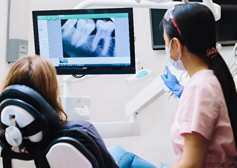
Digital Imaging
Radiographs are an invaluable tool in diagnosis and treatment planning. There are many guidelines that we follow. Radiographs allow us to see everything we cannot see with our own eyes. Radiographs enable us to detect cavities in between your teeth, determine bone level, and analyze the health of your bone. We can also examine the roots and nerves of teeth, diagnose lesions such as cysts or tumors, as well as assess damage when trauma occurs.
World Dental Specialities utilizes Digital Imaging Technologies within the office. Exposure time for digital dental radiographs is extremely minimal. With digital imaging, exposure time is about 50 percent less when compared to traditional radiographs. Digital imaging can also help us retrieve valuable diagnostic information. We may be able to see cavities better. Digital X rays of all our patients allows us to store patient images, and enables us to retrieve them easily whenever the need be.
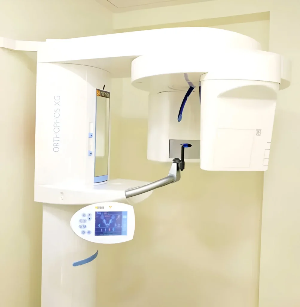
Cone Beam Computed Tomography (CBCT)
We, at World Dental Specialties have an inhouse CBCT machine which is the most advanced 3D IMAGING machine that offers rapid scan time and less radiation exposure compared to the CT technique. CBCT delivers three-dimensional volume rendering and offers accurate images with good spatial resolution.
Dental radiography is a vital part of clinical dentistry and CBCT (cone beam computed tomography), a fundamental, ever-evolving diagnostic tool that plays an integral role throughout the patient care pathway – allowing more accurate and informed diagnosis and treatment planning.
CBCT has revolutionized implant dentistry. Advancements in the technology have pioneered development in scanning equipment and innovations in viewing software allow for more precise treatment planning.
CBCT scanners vary in their capabilities; it is important to understand that achieving a high standard of diagnostic information is dependent on both the patient and the operator. As radiographers, we select the scanner, field of view and voxel parameters based on the clinical indications and individual patient. This optimizes the exposure and achieves the greatest level of diagnostic information.
CBCT’s undeniable advantage of multiplanar reconstruction has revolutionized implant dentistry; it allows visualization without the superimposition of structures. This ability to view structures from ‘different angles’ helps to more accurately evaluate the architecture and dimensions of the bone; the contour and visual density, the cortex and trabeculae pattern within the bone and the surrounding anatomical structures.
CBCT is also advantageous in assessing anatomy outside the dental alveolar region, including the zygoma, paranasal sinuses and airway.
Application Of CBCT In Dentistry
CBCT in Implantology:
CBCT is best because oral implants only require localized radiation exposure to the oral and maxillofacial region. CBCT scans help plan oral implants by measuring the distance between the alveolar crest and mandibular canal to prevent inferior alveolar nerve impingement, posterior lingual undercut perforation, and bone quality and density. They help plan maxilla oral implants, focusing on the nasopalatine canal and maxillary sinus. CBCT along with intraoral scanners help in precise implant planning on the computer and favilitates prcise implant placement with no error using a surgical guide which has revolutionised implant dentistry.
Digital implant planning and guided implant surgery offer many advantages in terms of optimised surgical and prosthetic treatment preparation and their predictable and successful implementation.
https://pubmed.ncbi.nlm.nih.gov
https://www.hindawi.com/journals/amse
CBCT in Endodontics:
CBCT provides a comprehensive description of the tooth’s morphology, including the specific characteristics such as the quantity and arrangement of canals, the dimensions and level of calcification of the pulp chamber, the orientation and curvature of the root, the presence of fractures, any anomalies resulting from dental procedures, and the extent of dental decay. Identifying outcomes associated with periradicular and periapical illness encompasses assessing root resorption levels and the presence of periapical osteolysis characteristics.
CBCT in Orthodontics:
CBCT offers a three-dimensional evaluation of craniofacial anatomy, maxillary transverse dimensions, and alveolar border conditions. In craniofacial orthodontics, CBCT can assess skeletal and soft tissues in all directions, maxillary expansion, and cleft evaluation. CBCT can better visualize 2D imaging findings like panoramic radiographs. This helps the orthodontist assess pathology in three dimensions and its link to the teeth.
CBCT in Periodontics:
Comparing intraoral periapical radiography and CBCT for detecting intrabony defects has demonstrated that the latter is significantly more accurate. Images from a CBCT can help better visualize periodontal tissue morphology. CBCT also provides high accuracy for identifying the involvement of the furcation. This method is beneficial for more advanced procedures.
CBCT in Pediatric Dentistry:
CBCT should only be used in pediatric patients when conventional radiography fails to offer significant information. CBCT pictures detect proximal carious lesions better than digital intraoral methods. CBCT should examine impacted supernumerary teeth to reduce danger to nearby anatomical tissues and cortical bone.
CBCT for TMJ Imaging:
CBCT is cheaper and safer than CT for TMJ evaluation. Assessing osseous TMJ problems is more advanced than older imaging techniques like radiography and MRI.
CBCT in Oral and Maxillofacial Surgery:
CBCT is routinely used to detect oral and maxillofacial cancers. Oral and maxillofacial fracture assessment and therapeutic planning prefer CBCT. It reduces the need for multiple 2D radiographs to find a similar thing. Benign jaw lesions have numerous radiological presentations. CBCT imaging determines the disease’s location, expansion, size, extent, and involvement of nearby vital structures.
Teeth in an Hour
Using a CBCT scan of your jaw structure and three-dimensional computer imaging technology, the exact placement of the implants is planned out before your appointment.
The implant surgery is virtually planned on the computer and surgical guide is fabricated which allows the position on the software to be transferred to the mouth.
A immediate temporary prosthesis on the implants is also planned on the software and then kept ready for placement on the day of the surgery.
The implants are placed in a precise position through the guide for optimum eathetics and function and the immediate long term temporary prosthesis is also delivered on that appointment.
Since so much time and effort is spent in the planning phase and it is so precise, the actual surgical appointment goes fairly quickly.
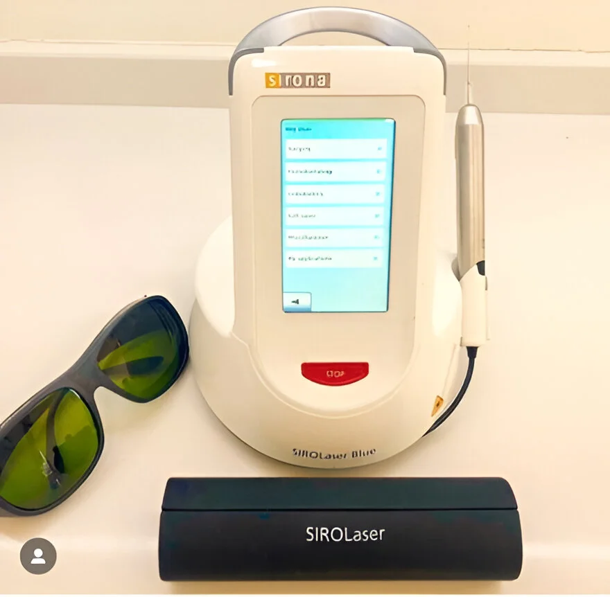
Dental Lasers
Lasers have advanced nearly every facet of medicine and we are now pleased to introduce them at World Dental Specialities for safe, fast, comfortable and effective procedures in all scopes of dentistry.
Why Lasers?
- Procedures performed using soft tissue dental lasers may not require sutures (stitches)
- Certain laser dentistry procedures do not require anesthesia.
- Laser dentistry minimizes bleeding because the high-energy light beam aids in the clotting (coagulation) of exposed blood vessels, thus inhibiting blood loss.
- Bacterial infections are minimized because the high-energy beam sterilizes the area being worked on.
- Damage to surrounding tissue is minimized.
- Wounds heal faster and tissues can be regenerated.
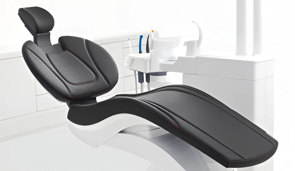
Dentsply Sirona Sinius Chairs
When it comes to optimizing operator access and patient comfort, Sinius chair makes no compromises. We pamper our patients with the Dentsply Sinius dental chair. Through the science of pressure mapping, the unique cushioning reduces pressure points and provides comfortable support across the patient’s entire body.
The chair boasts of ErgoMotion
- The combined motion of the backrest and seat prevents extension and compression of the spine & gives the patients a comfortable stress free experience
- The patient’s head always remains in the same position.
- There is no need to readjust the headrest when changing treatment position
It also has Lumbar support function
- The backrest can be individually adjusted to the patient by means of the lumbar support function in order to compensate for any anatomical differences and provide optimal support of the spine. It takes the shape of each patients back and thus limits the strain on the patients spine and lower back.
- Comfortably positioned patient
- Motion is also critical to patient comfort. The Dentsply Sinius dental chair synchronizes the chair movement with the natural motion of the patient. This ‘virtual pivot’ keeps the patient from having to readjust when the chair is lowered or raised.
Exceptional and unparalleled Highest hygienic standards (Purging function)
Sinius offers integrated hygiene function to prevent spread of infection. This purging function must be done after every patient visit to ensure infection free environment. Not only handpieces/threeway syringes get sanitized but the whole internal tubing system gets sterilized. After each patient, there is a cycle of 5 minutes of purging where suction hoses and the internal tubes get cleaned off from patients’ saliva and other infectious agents automatically, quickly and easily. At the end of each day, a 1 hour cycle is carried out which sanitizes the whole internal tubing system of the chair to reduce any retrograde infection from water lines.
- Integrated sanitization adapter: For regular flushing of the water lines and monthly sanitization, we can simply connect the instrument hoses to the integrated sanitization adapters and with the press of a button we have optimal water quality.
- Efficient suction hose cleaning: The suction hoses can also be cleaned automatically ‒ quickly and easily.
- Removable cuspidor: The swiveling cuspidor can be removed for easy cleaning.
- Elements can be thermally disinfected: Smooth, easy-to-clean surfaces. All hygiene-critical elements can be removed and sterilized or thermally disinfected.
- Automatic dosing: If desired, suction hose cleaning can also be performed with automatic addition of chemicals from the Dentsply Sirona maintenance and disinfectant list.
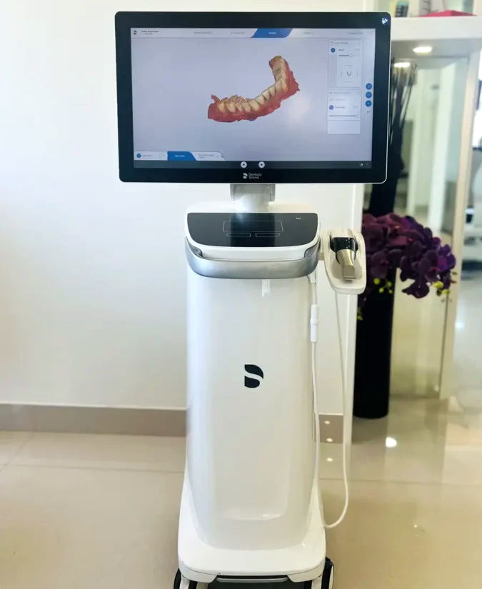
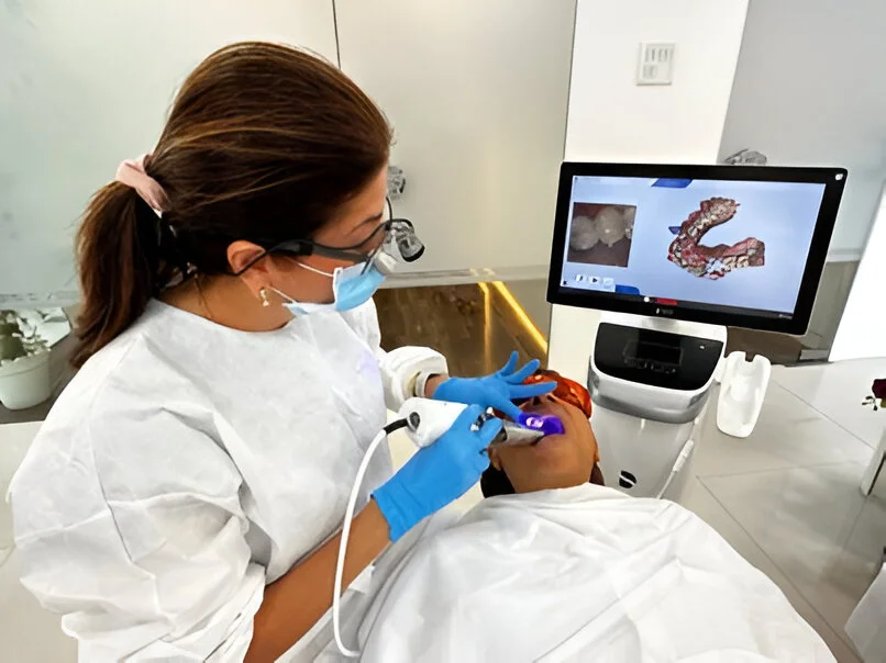
Intraoral Scanners
Intraoral scanners (IOS) are devices for capturing direct optical impressions in dentistry. Optical impressions reduce patient discomfort; IOS are time-efficient and simplify clinical procedures for the dentist, eliminating plaster models and allowing better communication with the dental technician and with patients
We at World Dental Specialities have always invested in cutting edge technology which enhances the treatment quality and patient experience and therefore have the best intraoral scanner in the world, Dentsply Sirona Primescan make patient care more comfortable and expand treatment workflows which allow quicker and more integrated treatment planning he advantages of intraoral scanners.
The advantages of the intraoral scanner as part of implant treatment include:
- Potential time-effectiveness: Since digital impression techniques are faster and don’t require dental impression materials and dental cast models, they can shorten the chair-side time.
- More accurate impressions: Impressions tend to deform, which can affect the fit of the final restoration. Impressions are poured in dental stone but there are always high chances of inaccuracies to occur in the production of dental cast models. The proficient use of intraoral scanners can help us mitigate the chances of distortion from the impression.
- Taking an impression before completion of osseointegration: Intraoral scans don’t require the use of conventional impression materials. This makes it possible to make an optical impression during surgery with minimal risk of contamination of the surgical field. In addition, intraoral scanners enable us to make an impression during the early stages of osseointegration without stressing the implant abutment component. https://blog.iti.org/clinical-insights/clinical-applications-and-advantages-of-intraoral-scanners-in-dental-implant-treatment/
- More comfortable for patients with a sensitive gag reflex and profuse salivation: Digital impression techniques can reduce foreign object sensation and breathing difficulties. In addition, impression materials need a setting time of about four minutes. Hence, digital impressions can be taken more comfortably than conventional impressions for patients. https://pubmed.ncbi.nlm.nih.gov
- Immediate evaluation of impressions and partial re-impression possible in the dentist’s chair: Intraoral scanners can immediately reconstruct the scanned image on the monitor and the area of insufficient scanning is marked clearly. In this case, it is possible to reproduce the area that was poorly scanned without losing all the impression data as with the conventional method.
- Easier to store and transfer digital images between the dental office and the laboratory. Since the patient’s impression is stored as digital data, the need to store multiple dental cast models is mitigated, which is excellent in terms of storage period and space. Moreover, transfer between the dental office and the dental laboratory is easier and faster.
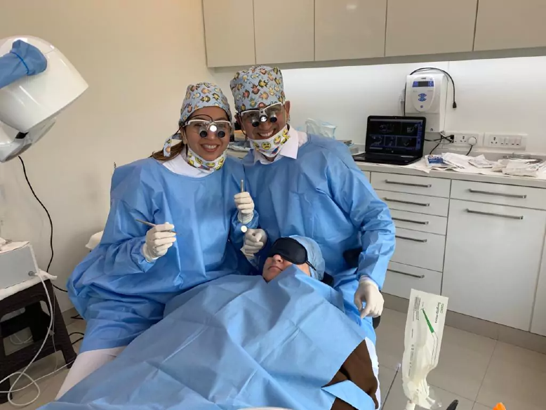
Magnification Loupes
It is often the biggest challenge during a procedure to be able to completely visualize what you are working on. Dental loupes enhance visual acuity as the dentists’ work involves small, detailed parts of the oral cavity which are located in a secluded and hard-to-reach area.
The advantages of using dental loupes
- Enhances visual acuity: Loupes allow the dentist to see more and ensure excellent end results.
- Improves precision and accuracy
- Can lead to more accurate diagnosis
- At World Dental Specialities, our Magnification loupes combined with a LED head light not only magnifies the area of interest, but also illuminates it for more precision-driven dentistry. Dental loupes in our armamentarium have significantly lifted the quality of dental care to a higher level.
In some cases, early signs of dental issues are unintentionally overlooked because of the naturally limited capacity of bare human eyes.
With the work area being magnified and illuminated, it becomes easier for us to visualise what’s going on inside the mouth, allowing to see things that can’t be seen with bare eyes like an almost invisible stain or a very minor crack on the teeth too.
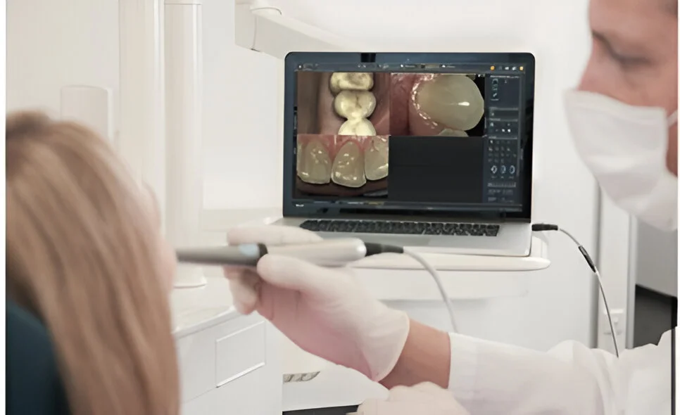
Intra Oral Cameras
This is probably one of the most useful tools in our armamentarium for our New Patient examinations. We at World Dental Specialities have the latest Dentsply Sirona SiroCam UAF Plus ,which is the one of the best intra oral cameras. It provides clear case visualization, thus enabling patients to understand diagnosis and the recommended treatment plan, improving overall communication and patient experience.
Convenient Diagnosis
A dentist can easily detect dental problems without the patient keeping the mouth open for a few hours by means of an intraoral camera to explore the oral cavity. This great progress has made dental appointments smoother and more convenient for patients.
Preventive Diagnosis and Care
Our Intraoral camera is not only less invasive, but helps to detect oral health issues right at the commencement stage. It can be used to identify possible issues with dental health that the naked eye cannot detect. In fact, the early detection of an oral problem avoids significant damage to your teeth and prevents future complications.
More Transparent Patient Education
Dentists often find it difficult to communicate their problems and the device can be used to communicate with the patients better. They can be used to help patients to consider their teeth’s condition over and above the diagnostic benefits.